In Vivo Confocal Microscopy (IVCM)
A practical guide to interpreting corneal nerve structure, immune activity, and pathological patterns. Focused on real-world findings such as nerve loss, microneuromas, and inflammation markers.
1. What is Confocal Microscopy?
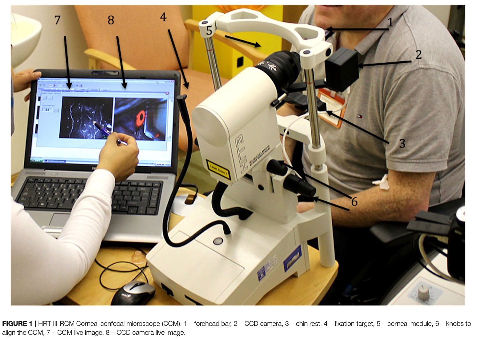
In vivo confocal microscopy (IVCM) is a high-resolution imaging method that allows visualization of corneal nerves, immune cells, and epithelial structure at near-cellular resolution. It is commonly used in neuropathic corneal pain and dry eye disease evaluation.
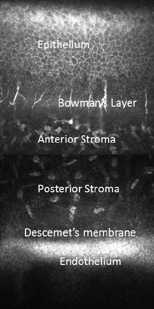
The human cornea is a transparent, avascular tissue that serves as the eye's outermost layer. It is composed of five distinct layers, each with unique structural and functional properties:
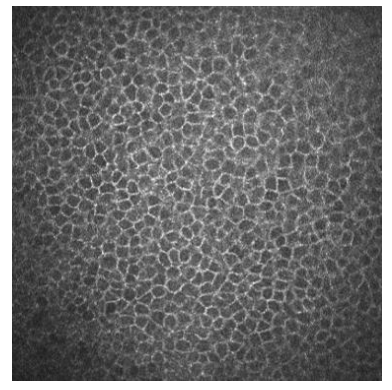
- Corneal Epithelium: ~ 50 μm deep. This outer, fast-regenerating layer is 5 to 7 cells thick and makes up roughly 10% of the total corneal thickness.
- Bowman's Layer (Membrane): ~ 8–14 μm deep. A very thin, dense, and tough fibrous sheet of connective tissue that protects the underlying stroma.
- Stroma (Substantia Propria): ~ 400–500 μm deep. The thickest layer, constituting roughly 80–90% of the total corneal depth. It is composed of precisely arranged collagen fibers that provide structural strength and transparency.
- Descemet's Membrane: ~ 5–15 μm deep (thickens with age). A thin, elastic basal lamina that separates the stroma from the inner endothelium.
- Endothelium: ~ 4–10 μm deep. A single, specialized layer of cells on the innermost surface that pumps fluid out of the stroma to keep the cornea clear.
Typical Epithelium:
Typical Subbasal Nerve Plexus:
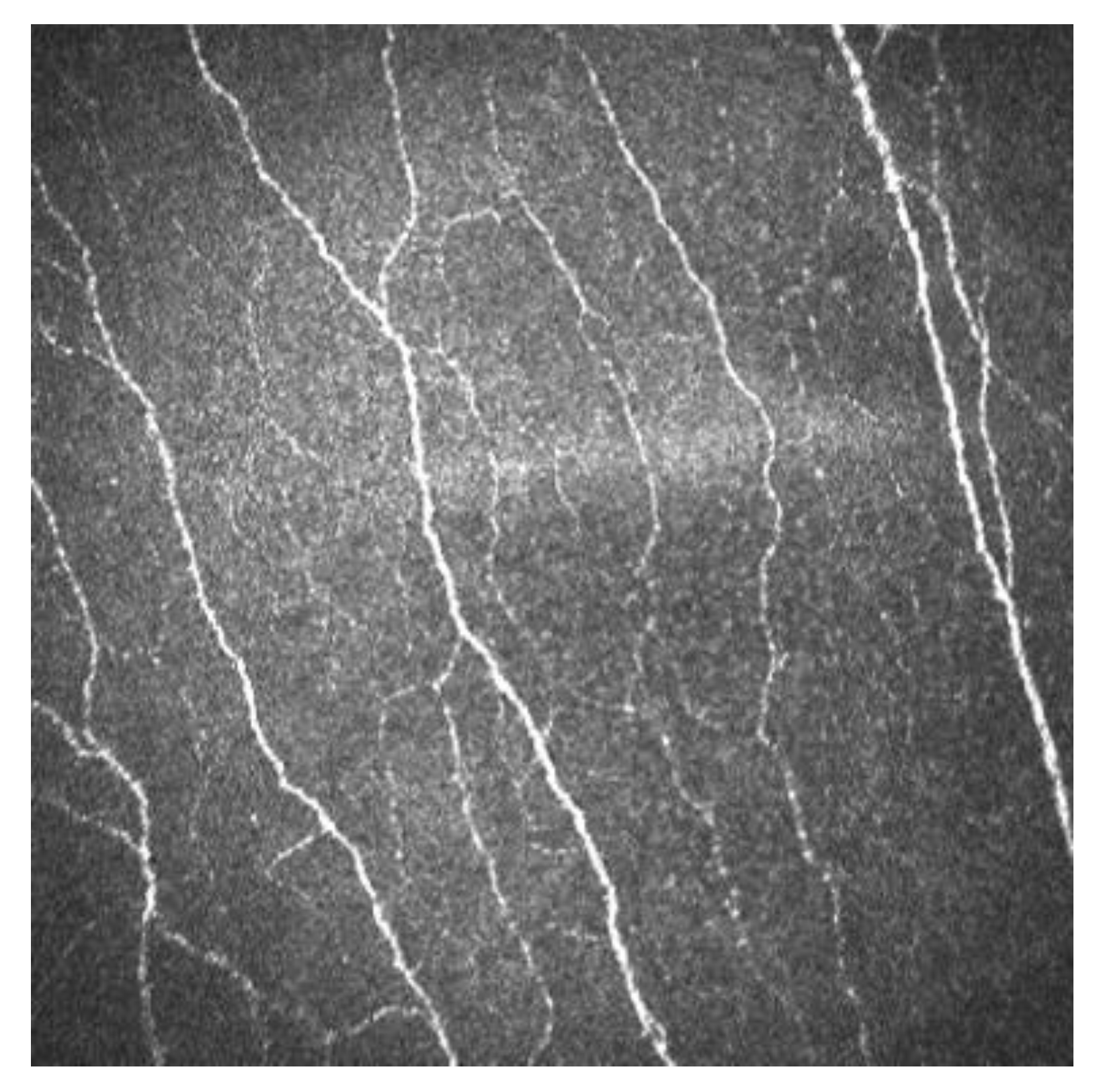
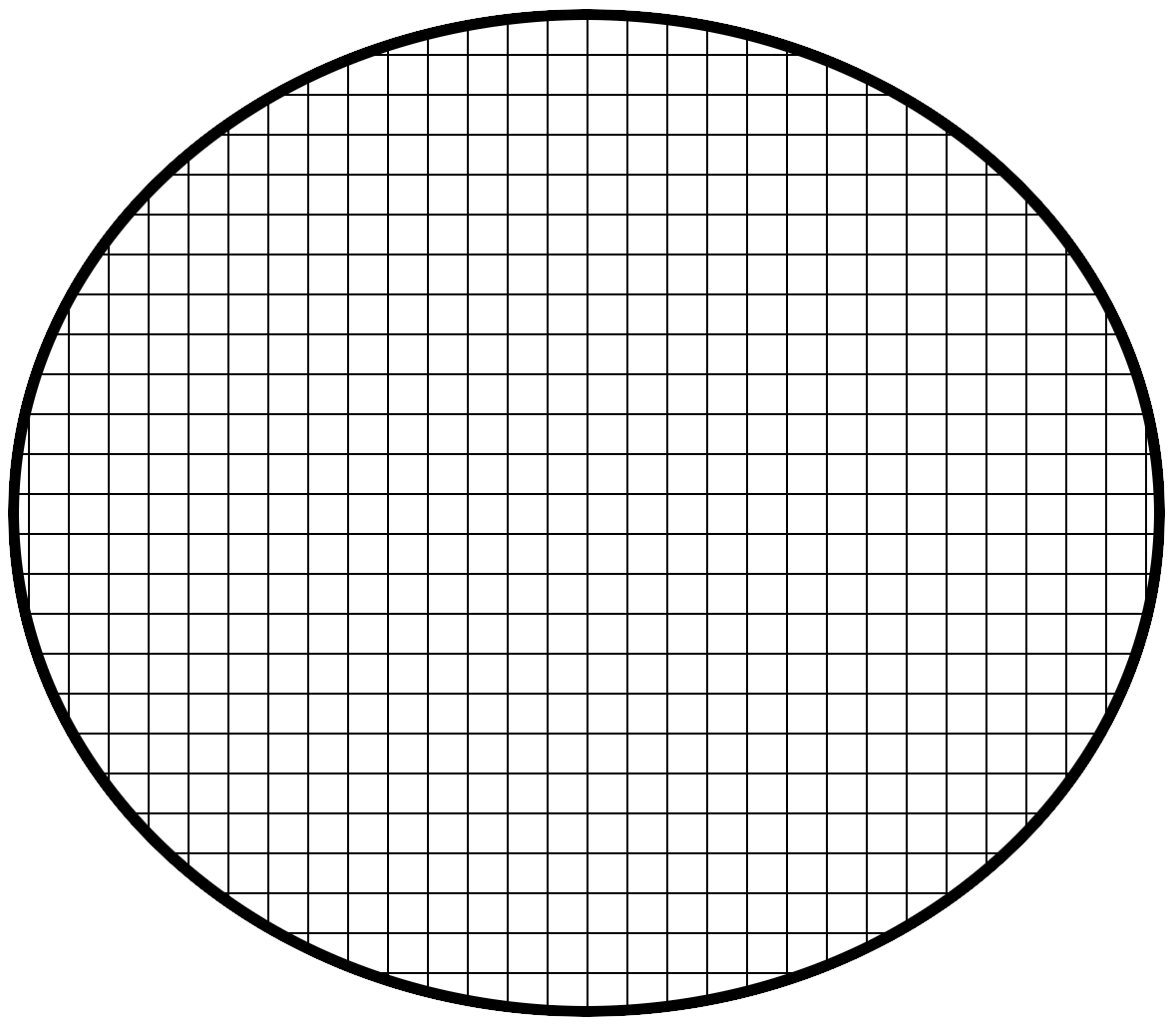
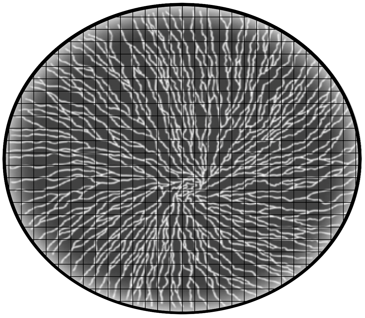
Typical Field of View of IVCM is 400 μm x 400 μm (0.16 mm²) which corresponds to a small fraction of the corneal surface area. The following image shows the scale of the cornea and the field of view of IVCM.